
Cancer diagnosis has moved far beyond the traditional biopsy and basic imaging. Today’s oncology departments combine molecular testing, functional imaging, and computational analysis to detect malignancies earlier, classify them more precisely, and tailor treatment to the individual patient. These diagnostic advances matter because they directly influence survival rates, treatment toxicity, and quality of life during and after cancer care.
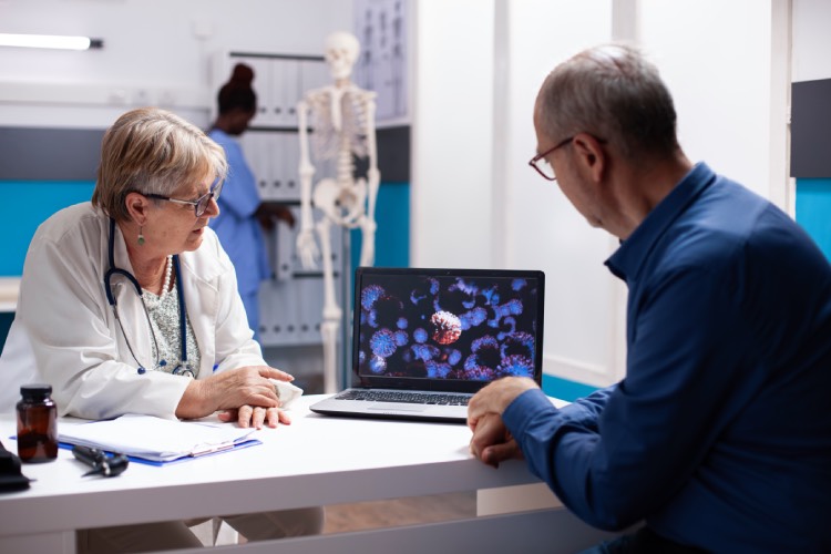
The shift toward precision diagnostics reflects a fundamental change in how we understand cancer itself. Rather than viewing breast cancer or lung cancer as single diseases, modern pathology recognises dozens of subtypes within each category, each responding differently to chemotherapy, immunotherapy, or targeted drugs. Getting the diagnosis right at the molecular level determines whether a patient receives the most effective treatment or wastes months on therapies unlikely to work.
The Role of Molecular and Genetic Testing
Molecular diagnostics examine the genetic mutations, protein expressions, and chromosomal changes that drive cancer growth. A tumour biopsy now routinely undergoes next-generation sequencing to identify actionable mutations in genes like EGFR, BRAF, or BRCA. These findings determine eligibility for targeted therapies that block specific pathways cancer cells use to multiply.
Immunohistochemistry testing measures protein markers on tumour cells. In breast cancer, testing for oestrogen receptor, progesterone receptor, and HER2 status separates patients into treatment groups with vastly different prognoses and drug regimens. A HER2-positive tumour receives drugs like trastuzumab, while a triple-negative tumour requires chemotherapy or immunotherapy combinations.
Liquid biopsies represent an emerging frontier. These blood tests detect circulating tumour DNA shed by cancer cells, offering a non-invasive method to monitor treatment response or detect recurrence months before conventional imaging would show new growth. The technology remains most useful in patients with known cancer rather than as a general screening tool, but refinements continue.
Advanced Imaging Technologies in Oncology
PET-CT scans combine metabolic and anatomical imaging to show not just where a mass sits but how active it is. Cancer cells consume glucose at higher rates than normal tissue, lighting up on PET scans and revealing metastases too small or too metabolically similar to surrounding tissue for CT alone to catch. This technique proves particularly valuable in lymphoma staging, where treatment intensity depends on disease spread, and in assessing treatment response midway through chemotherapy.
MRI sequences tailored to specific cancer types provide detail that CT cannot match. Diffusion-weighted imaging measures how water moves through tissue, helping distinguish benign from malignant liver lesions or detect prostate cancer within the gland. Contrast-enhanced MRI maps blood flow patterns that reveal tumour neovasculature, the chaotic new blood vessels that feed growing cancers.
Artificial intelligence now assists radiologists in interpreting these scans. Algorithms trained on thousands of images highlight suspicious nodules, measure tumour volumes with precision humans struggle to match, and track subtle changes between sequential scans. Machine learning applications in healthcare extend beyond imaging to risk prediction and treatment planning, though radiologists remain essential for contextualising findings within each patient’s clinical picture.
Multidisciplinary Tumour Boards and Integrated Diagnostics
Complex cancer cases benefit from collective expertise. Tumour boards bring together medical oncologists, radiation oncologists, surgeons, radiologists, and pathologists to review diagnostic findings and debate treatment options. A pancreatic cancer case might hinge on whether imaging shows the tumour encasing a major blood vessel, rendering surgery inadvisable, or whether molecular testing reveals a rare subtype responsive to specific chemotherapy.
These collaborative discussions prevent the fragmentation that occurs when specialists work in isolation. A surgeon might propose resection based on imaging, while the pathologist’s review reveals aggressive histology warranting neoadjuvant chemotherapy first. Radiation oncologists contribute when anatomical constraints make surgery high-risk but focused radiation could achieve local control. The patient receives a unified plan rather than conflicting opinions.
Specialised Cancer Care in the UAE
Comprehensive cancer centres integrate diagnostic and treatment services under one roof, reducing the coordination burden on patients navigating multiple appointments. Facilities in Abu Dhabi have developed subspecialised units focusing on specific cancer types, allowing clinicians to concentrate expertise and stay current with rapidly evolving treatment protocols. Patients with hepatobiliary cancers—those affecting the liver, bile ducts, and pancreas—require particularly specialised care given the complex anatomy and challenging surgical access. Hepatopancreatobiliary treatment Abu Dhabi centres offer multidisciplinary teams experienced in these technically demanding cases, combining advanced imaging for surgical planning with molecular testing to identify patients who might benefit from targeted therapy or immunotherapy before or after surgery.
The regional concentration of expertise matters because hepatobiliary cancers present diagnostic challenges. Distinguishing between benign bile duct strictures and cholangiocarcinoma requires expert interpretation of imaging alongside brush cytology and sometimes endoscopic ultrasound. Treatment decisions balance aggressive surgery with substantial recovery periods against the reality that some cancers have spread microscopically beyond what imaging reveals.
The Global Cancer Burden and Diagnostic Priorities
Cancer remains a leading cause of death worldwide, with nearly 10 million deaths in 2020 according to global health statistics. Lung, colorectal, liver, stomach, and breast cancers account for the majority of cancer mortality. Early detection programmes dramatically improve outcomes for several of these cancers, but only when diagnostic infrastructure and trained personnel exist to act on screening results.
Low-resource settings face different diagnostic challenges than high-income healthcare systems. A sophisticated PET-CT scanner provides limited benefit if pathology labs cannot perform immunohistochemistry or if drug supply chains cannot deliver the targeted therapies that molecular testing identifies. Effective cancer control requires matching diagnostic capabilities to available treatments and building the entire care pathway, not just acquiring advanced equipment.
Practical Considerations for Patients
Understanding your diagnostic workup helps you participate in treatment decisions. Ask which specific tests the oncology team plans and what each reveals about your cancer. Molecular testing takes one to three weeks for results, delaying treatment start but potentially identifying therapies with better response rates and fewer side effects than standard chemotherapy.
Request copies of pathology and radiology reports. These documents contain details your oncologist may not mention in conversation but which matter if you seek a second opinion or transfer care. Staging information, margin status from surgical specimens, and specific mutations detected all influence subsequent treatment recommendations.
Second opinions make sense for rare cancers, when initial recommendations seem aggressive or conservative relative to your research, or when treatment would significantly impact fertility, organ function, or quality of life. Bring all imaging studies on disc rather than just reports, as expert radiologists often identify details the original reading missed or interpreted differently.
Looking Ahead
Diagnostic technology continues advancing faster than clinical trials can validate every application. Circulating tumour cells, methylation patterns, and proteomic signatures all show promise but require more evidence before becoming standard practice. The challenge lies in determining which innovations genuinely improve patient outcomes versus those that add cost and complexity without changing treatment decisions or survival.
Precision diagnostics work best when integrated with precision treatment. Identifying a druggable mutation means little if the corresponding drug costs prohibitively or causes toxicity the patient cannot tolerate. The goal remains matching each patient to the treatment most likely to control their cancer with acceptable side effects, using diagnostics as the tool to make that match rather than as an end in themselves.


Leave a Reply